Mammalian Kidney Dissection


Suggested Time

Time45–90 min
Span1–2 Class Periods
Ideal Group Size
2 students per group
Safety Supplies (Required)


Safety Supplies (Recommended)

 View
ViewRecommended Dissecton Supplies
Kidney Dissection Video
Kidney External Anatomy
- Place the preserved pig kidney on your dissecting tray.
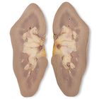
- Observe and feel the renal capsule (Figure 1). This structure encapsulates the kidney and is made up of dense, irregular connective tissue. It provides protection and helps maintain the shape of the kidney.
- Locate the hilus (Figures 1 and 2). This is an indentation where the ureter exits and blood vessels either enter or exit the kidney.
- The ureter is located within the adipose tissue that is attached to the hilus. It drains urine from the renal pelvis into the bladder.
- The renal artery and renal vein are also found in the adipose tissue of the hilus. The renal artery is located directly above the renal vein and has a slightly thicker wall. The renal artery supplies the kidney with blood, carrying water and wastes to be filtered and removed. After the blood has been filtered, many veins throughout the capsule converge into the renal vein, which returns the filtered blood to the body.
- Remove any adipose tissue attached to the renal capsule and hilus. Consult Figure 2 to ensure that you do not remove too much of the tissue around the ureter and the renal artery and vein.

Kidney Internal Anatomy
- Carefully make a lateral, longitudinal cut through the kidney and lay the 2 halves on the dissecting tray.
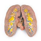
- Locate the cortex and medulla. The medulla lies below the cortex. Observe and record the appearance of each region.
- Look closely at the cortex section of the kidney. Locate the collecting ducts and follow them into the renal medulla (Figure 3). Urine is formed in microscopic structures called nephrons and then passed to the collecting ducts.
- Locate the conical renal pyramids in the renal medulla (Figure 4). The base of each renal pyramid lies next to the cortex, while the tip forms the renal papilla. Urine in the collecting ducts travel through the renal pyramids to the renal papillae.
- Adjacent to the renal pyramids are the renal columns, that expand from the cortex through the medulla, composed of cortical tissue that has a granular texture similar to that of the cortex (Figure 4). The renal cortex, renal pyramids, and renal columns collectively are called the renal lobe. Urine passes through the renal lobe and is discharged through the renal papilla, the apex of the renal pyramids, into the minor calyx.
- Locate a minor calyx, major calyx, and the renal pelvis. (Figure 5). Urine from a renal papilla empties into a minor calyx. Minor calyces merge to form major calyces. Major calyces merge to form the renal pelvis.
- Trace a path through the minor and major calyces and into the renal pelvis. Urine from the renal pelvis then drains urine into the ureter where it is transferred to the bladder.
- After you have observed all the structures of the kidney, dispose of the specimen in accordance with local guidelines and your teacher’s instructions.

Figure 3

Figure 4

Figure 5