
Earthworm Dissection

Suggested Time

Time45 minutes
Span1 Class Period
Ideal Group Size
2 students per group
Safety Supplies (Required)


Safety Supplies (Recommended)

 View
ViewRecommended Specimen(s)
Recommended Dissecton Supplies
General Dissection Resources
Videos
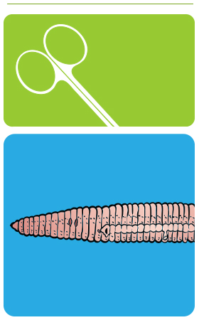
External Earthworm Anatomy

- First, identify the anterior and posterior ends. The anterior end is somewhat larger than the posterior. The earthworm has no head and no appendages, but it does possess external characteristics to study.
- Locate the mouth opening and the prostomium at the anterior end.
- Earthworms are annelids, or segmented worms that have bodies made up of a series of ring-like segments. Count and record the number of segments. There may be as many as 100.
- Observe each segment closely.
- Locate the tiny bristles on the ventral surface called setae. These help the worm move through soil.
- Each segment also contains a pair of small excretory pores called nephridiopores. You may need to use a hand lens or stereomicroscope to see these openings.
- Earthworms are hermaphroditic so both sex organs are present. Find the openings to the oviducts, which release eggs at segment 14, counting from the anterior end. Also locate the openings to the sperm ducts on segment 15.
- The clitellum is the enlarged structure that begins at segment 31. It secretes mucus that holds 2 earthworms together during mating and produces a cocoon in which eggs and sperm are deposited for external fertilization.
Internal Earthworm Anatomy

- Place the earthworm on its ventral side. (The ventral side is more flattened than the dorsal side.) Using dissecting scissors, cut through the dorsal body wall posterior to the clitellum and continue the incision toward the prostomium. Be careful not to cut too deep or you will slice into the digestive system.
- Using forceps, spread the incision open and pin the body wall to the dissecting pan as shown in the illustration.
- Identify the thin walls between each segment. These are called septa.
- Using the diagram above, identify the organs of the digestive system:
- Pharynx
- Esophagus
- Crop (stores food)
- Gizzard (grinds food)
- Intestine
- Remove part of the intestine posterior to the gizzard and locate the ventral blood vessel.
- Identify the 5 pairs of aortic arches, or hearts, which circle the esophagus.
- Identify the cerebral ganglia and the ventral nerve cord that extends from them, down the ventral body wall.
- Locate the excretory organs called nephridia in each segment. Nephridia remove nitrogenous waste.
- Also locate the seminal receptacles and seminal vesicles.
- Once you have observed the structures of the earthworm, dispose of the specimen in accordance with local guidelines and your teacher’s instructions.