
My Cart
Your Shopping Cart is currently empty. Use Quick Order or Search to quickly add items to your order!
This lab introduces students to the variation found in the fungal phylum Ascomycota. Students examine four representatives: Anthracobia muelleri, Eurotium chevalieri, Schizosaccharomyces octosporus, and Sordaria fimicola. Schizosaccharomyces is a unicellular yeast, while the others show typical mycelial growth.
Ascomycete Fruiting Set (155822)
Bunsen burners or alcohol lamps
beakers or cups for water
This activity requires that students work with fungal cultures and open flames. Have students wipe down all work surfaces before and after the lab, using disinfectant. Ensure that students wash their hands before and after the lab. Destroy any cultures remaining at the end of the lab by autoclaving or by flooding them with disinfectant overnight before proper disposal.
Ensure that students understand and adhere to safe laboratory practices when performing any activity in the classroom or lab. Demonstrate the protocol for correctly using the instruments and materials necessary to complete the activities, and emphasize the importance of proper usage. Use personal protective equipment such as safety glasses or goggles, gloves, and aprons when appropriate. Model proper laboratory safety practices for your students and require them to adhere to all laboratory safety rules.
Students may work individually or in pairs.
Scheduling presents a problem because the cultures must be used when ascocarps (fruiting bodies) are at their optimum stage. A plate that is ready to use can be maintained under refrigeration for up to 2 days before use.
Anthracobia
Look for circular growths of about 1 mm diameter that develop near the margin of the plate. These are the apothecia.
Eurotium
Use the plate when there are large green areas, produced by clusters of conidia. If you delay observation, the spores will be discharged.
Schizosaccharomyces
This plate can be used at almost any time.
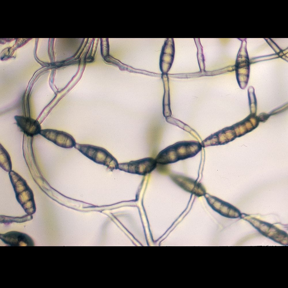
Sordaria
Watch for the development of dark specks (usually appearing first near the margin of the plate). These are the perithecia, which contain clusters of asci. If you wait too long after this point, the asci will have opened, discharging their spores.
Refer to the information about ascomycetes in the Techniques for Studying Bacteria and Fungi manual shipped with your order. (The manual can also be downloaded from carolina.com under Teacher Resources.)
Set up a workstation for each plate culture—Anthracobia, Eurotium, Schizosaccharomyces, and Sordaria—with the following:
plate culture
dropping pipet in beaker of water
dissecting needle
microscope slides
Bunsen burner or alcohol lamp
coverslips
compound microscope (with scanning objective for 40× viewing)
Observe your slide using the scanning and low-power lenses of a microscope. The ascocarps are flask-shaped. Some will be crushed, releasing asci, which are tubular. Look closely at the end of an ascus and you may see the pore through which the spores will be ejected. Count the spores in an ascus. What is the normal number of spores per ascus? 8 These spores were produced by meiosis, and meiosis results in a tetrad of spores. What do you think happened after meiosis to produce the number that you observe?
Each spore underwent mitosis, to produce a total of eight ascospores.
Below is a description of three basic types of ascocarps. Using your observations, match each culture with its ascocarp.
| Ascocarp type | Description | Example |
| Apothecium | An open ascocarp that does not enclose the asci. | Anthracobia |
| Cleistothecium | A completely closed ascocarp. | Eurotium |
| Perithecium | An ascocarp with one opening. | Sordaria |