Products Suggestions:

Products suggessions:

We use cookies to provide you with a great user experience. By using our site, you accept our use of cookies . You can review our cookie and privacy policy here.

Modeling Mitosis and Meiosis

By Candace Berkeley
Product Developer

Lily pollen meiosis

Have student groups use sock "chromosomes" and string to model mitosis and meiosis. These 2 simple activities take approximately 30 min to complete. Discussion questions and answers are provided so that you can assess student understanding of each type of cell division.

National Science Education Standards

This activity is appropriate for high school students and addresses the following National Science Education Standards:

Grades 9–12

Life Science

  • The Cell
  • The Molecular Basis of Heredity

Materials

  • Socks
  • String or Yarn
  • Scissors
  • Measuring Tape(s)

Preparation

>
  • Divide the class into groups of 4.
  • Provide each group with the following materials:
    • 2 pairs of tube socks and 2 pairs of ankle socks
    • Approximately 60 feet (18.25 m) of string
    • Scissors
  • Decide how you want to handle the discussion questions (e.g., treat them literally as discussion, paste them into a photocopy master for homework or classwork, supply them to students electronically).

Procedure

Modeling Mitosis

  1. Tie the ends of a 10-ft (3-m) piece of string together and form a circle on the table. This represents the cell membrane of a parent cell.
  2. Make a smaller circle from a 5-ft (1.5-m) piece of string to create a nuclear membrane, and place it inside the cell membrane.
  3. Place 1 sock of each pair into its mate and jumble the socks inside the nuclear membrane.
  4. During interphase, the cell prepares for nuclear division and DNA replicates. To simulate replication, remove a mate from each pair of socks. Match sister chromatids and connect them at their centromere by tying the matching pairs together at their centers with a small piece of string. Place them back into the nuclear membrane, jumbling them up.
  5. Mitotic division now begins with prophase. During this phase, DNA condenses into chromosomes made up of 2 identical sister chromatids, and the nuclear membrane breaks down. To model prophase, remove the nuclear membrane and evenly distribute the pairs of tied-together socks inside the cell membrane.
  6. During metaphase, replicated chromosomes line up along the equator of the cell. Simulate this using the sock chromosomes.
  7. During anaphase, sister chromatids are pulled toward opposite sides of the cell. Once chromatids are pulled apart, they are now unreplicated chromosomes. Simulate anaphase by untying the sister chromatids and pulling them toward opposite sides of the cell.
  8. Telophase, the last phase of mitotic division, and cytokinesis, the division of the cytoplasm, often occur simultaneously. Cluster the unreplicated chromosomes at each side of the cell and use strings (about 3 ft [1 m]) to create a new nuclear membrane around each of the 2 clusters. Pinch the cell membrane string together until it meets between the 2 new nuclei. Cut it and tie the ends. Now you have 2 daughter cells, each with its own nucleus.

Modeling Meiosis

  1. Tie the ends of a piece of 10-ft (3-m) string together and form a circle on the table. This represents the cell membrane of a parent cell.
  2. Make a smaller circle from a 5-ft (1.5-m) piece of string to create a nuclear membrane, and place it inside the cell membrane.
  3. Place 1 sock of each pair into its mate and jumble the socks inside the nuclear membrane. The 2 tube sock chromosomes represent a homologous pair. One chromosome was received from the mother, and 1 was received from the father. This is also true for the ankle sock chromosomes.
  4. During interphase, prior to meiosis I, the cell prepares for nuclear division and DNA replicates. To simulate replication, remove the mate of each pair of socks. Match sister chromatids and connect them at the centromere by tying the matching pairs together at their centers with a small piece of string. Place them back into the nuclear membrane and jumble them.
  5. To simulate prophase I, remove the nuclear membrane and match homologous pairs of chromosomes together near the center of the cell.
  6. Model metaphase I by lining the homologous pairs of chromosomes along the cell’s equator.
  7. Pull homologous chromosomes apart (toward opposite sides of the cell) to represent anaphase I.
  8. To simulate telophase I and cytokinesis, cluster each batch of separated chromosomes together on its side of the cell and create a new nuclear membrane around each of the 2 clusters. Pinch the cell membrane together between the 2 nuclei, cut it, and tie the ends to make 2 daughter cells. Note: To have enough working space for the remainder of meiosis, you may need to switch to longer strings for the membranes of these 2 daughter cells—perhaps 8- to 10-ft (2.4- to 3-m) lengths.
  9. Prophase II follows cytokinesis. DNA is not replicated before prophase II. Model this phase by removing the nuclear membranes of each cell and spreading out the chromosomes inside the cell.
  10. Simulate metaphase II by lining the chromosomes along the equator of each cell.
  11. In anaphase II, sister chromatids are separated. Untie the sister chromatids and pull them toward opposite sides of the cell.
  12. During telophase II and cytokinesis, chromosomes bunch together at opposite ends of the cells, new nuclear membranes form, and the cytoplasm divides. Model these phases of the cell cycle. At the end of meiosis II, 4 daughter cells are present.

Follow-up

Students may struggle with understanding the difference between chromosomes, chromatids, and homologous pairs as they answer the discussion questions. Take a few minutes to discuss the differences between these terms as a class, using models or drawings to help. As with any scientific model, be sure to discuss the model’s limitations so that students do not develop misconceptions. For example, discuss mechanisms that prevent cells from becoming smaller as they divide (e.g., cells expand before division). String lengths and classroom space probably prevent modeling the proportions more accurately. Make sure that students are aware that the pinching of the cell membrane is characteristic of animal cell cytokinesis but that the process differs in plant cells.

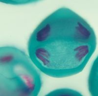
As an extension of this activity, have students observe cells in various stages of mitotic and/or meiotic division. Consider using the Fish and Onion Mitosis Slide Set (item #308816) or the Mitosis and Meiosis Slide Set (item #308826).

Discussion questions and answers

Modeling Mitosis

  1. In interphase, how many chromosomes were present in the parent cell before replication and after replication?
    Four chromosomes were present before and after replication in interphase. (There were 8 chromatids present after replication.)
  2. How many chromosomes were present in each daughter cell? Are they genetically identical? Explain.
    Four chromosomes were present in each daughter cell. They are genetically identical. This is because the chromosomes that were initially present were replicated and joined at the centromere. Identical chromatids were then split apart, resulting in genetically identical daughter cells.
  3. In what areas of your body do you think mitosis occurs regularly? Give specific examples.
    Mitosis occurs regularly in cells that are frequently damaged or lost. Skin cells, cheek cells, and cells lining the digestive tract are examples of cells that undergo regular mitotic division.

Modeling Meiosis

  1. In interphase, how many chromosomes were present in the parent cell before replication and after replication? How many homologous pairs were present?
    Four chromosomes were present in the parent cell before and after replication. Two homologous pairs were present.
  2. After meiosis I, how many chromosomes were present in each of the 2 daughter cells? Were homologous pairs present in the new daughter cells?
    Two chromosomes were in each daughter cell. Homologous pairs were not present in the daughter cells.
  3. After meiosis II, how many chromosomes were present in each of the 4 daughter cells?
    Two chromosomes were in each of the four daughter cells.
  4. What type of cell is formed by meiosis? Why is it important for the daughter cells to have half the number of chromosomes as the parent cell?
    Meiosis forms sex cells (also called gametes, or sperm and egg cells). It is important that these cells have half the number of chromosomes as the parent cell because during fertilization an egg and a sperm cell merge, doubling the number of chromosomes in the resulting cell. This allows formation of a zygote with the number of chromosomes needed for development of the embryo.

Visit our Mitosis page for more products and resources.